
Fully Automated Quantification Of LV Regional Wall Motion From Echocardiograms To Detect Myocardial Infarction.
Shan Gao, Mihaela Porumb, Angela Mumith, Andrew Parker, Seamus Walker, Gaynor Jones, Agis Chartsias, Jorge Oliveira, Will Hawkes, Rizwan Sarwar, Paul Leeson, Arian Beqiri.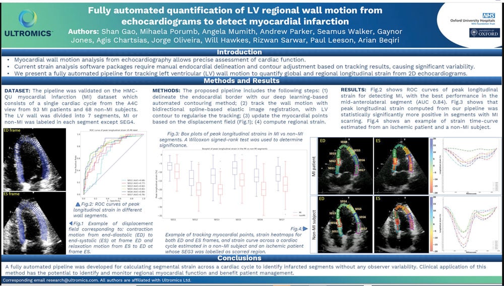
Background
Myocardial wall motion analysis from echocardiography allows precise assessment of cardiac function. Current strain analysis software packages require manual endocardial delineation and contour adjustment based on tracking results, causing significant variability.
We present a fully automated pipeline for tracking left ventricular (LV) wall motion to quantify global and regional longitudinal strain from 2D echocardiograms.
Dataset
The pipeline was validated on the HMC-QU myocardial infarction (MI) dataset which consists of a single cardiac cycle from the A4C view from 93 MI patients and 68 non-MI subjects. The LV wall was divided into 7 segments, MI or non-MI was labeled in each segment except SEG4.
Methods
The proposed pipeline includes the following steps: (1) delineate the endocardial border with our deep learning-based automated contouring method; (2) track the wall motion with bidirectional spline-based elastic image registration, with LV contour to regularise the tracking; (3) update the myocardial points based on the displacement field (Fig.1); (4) compute regional strain.
Results
Fig.2 shows ROC curves of peak longitudinal strain for detecting MI, with the best performance in the mid-anterolateral segment (AUC 0.84). Fig.3 shows that peak longitudinal strain computed from our pipeline was statistically significantly more positive in segments with MI scarring. Fig.4 shows an example of strain time-curve estimated from an ischemic patient and a non-MI subject.
Conclusion
A fully automated pipeline was developed for calculating segmental strain across a cardiac cycle to identify infarcted segments without any observer variability. Clinical application of this method has the potential to identify and monitor regional myocardial function and benefit patient management.