In this post
Akerman, A.P., Porumb, M., Scott, C. G., Beqiri, A., Chartsias, A., Ryu, A.J., Hawkes, W., Huntley, G.D., Arystan , A.Z., Kane, G.C., Pislaru, S.V., Lopez Jiminez , F., Sarwar, R., O’Driscoll., Leeson, P., Upton, R., Woodward, G., Pellikka, P.A.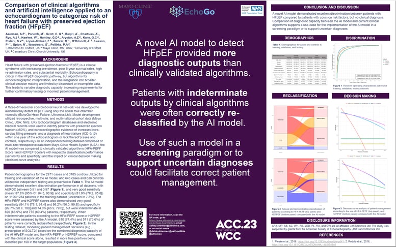
Background
Heart failure with preserved ejection fraction (HFpEF) is a clinical syndrome with increasing prevalence, poor 5 year survival rates, high re admission rates, and substantial morbidity. Echocardiography is critical in the HFpEF diagnostic pathway, but algorithms for echocardiographic interpretation, and the integration into broader clinical decision making are limited by discordant or incomplete data. This leads to variable diagnostic capacity, increasing requirements for further confirmatory testing or incorrect patient management.
Methods
A three dimensional convolutional neural network was developed to automatically detect HFpEF using only the apical four chamber videoclip (EchoGo Heart Failure; Ultromics Ltd). Model development utilized retrospective, multi site, and multi national cohort data (Mayo Clinic, USA; NHS, UK).
Echocardiogram databases and electronic medical records were used to identify patients with preserved ejection fraction (≥50%), and echocardiographic evidence of increased intra cardiac filling pressure, and a diagnosis of heart failure (ICD 9/10) within one year of the echocardiogram or lack thereof (cases and controls, respectively).
In an independent testing dataset comprised of multi site retrospective data from Mayo Clinic Health System ( USA), the AI model was compared to clinically validated algorithms (HFA PEFF Score 1 and H2FPEF Score 2 ) with respect to classification performance (sensitivity and specificity) and the impact on clinical decision making (decision curve analysis).

EchoGo® Heart Failure can accurately detect HFpEF using only a single transthoracic echocardiography videoclip. Click here to learn more about the clinical breakthrough in HFpEF detection.
Results
Patient demographics for the 2971 cases and 3785 controls utilized for training and validation of the AI model, and 646 cases and 638 controls utilized for independent testing are presented in Table 1 .
The AI model demonstrated excellent discrimination performance in all datasets, with AUROC between 0.91 and 0.97 ( Figure 1 ), and very good sensitivity (mean: 87.8% [95% CI: 84.5, 90.9]) and specificity (81.9% [78.2, 85.6]) on 1190/1284 patients in the training dataset (uncertain in 7.3%).
The HFA PEFF and H2FPEF scores also demonstrated very good sensitivity (84.1% [78.1, 91.4] and 98.2% [96.3, 99.8]) and specificity (99.7% [98.8, 100] and 74.0% [66.9, 79.0]), but were indeterminate in 820 (63.9%) and 776 (60.4%) patients, respectively.
When indeterminate patients according to the HFA PEFF score or H2FPEF score were assessed by the AI model, 610 (74.4%) and 571 (73.6%) of patients were correctly reclassified (respectively; Figure 2 ).
In the testing dataset, modelling patient management decisions (e.g., prescription of SGLT2i) based on the combined diagnostic capacity of the AI HFpEF model and the HFA PEFF or H2FPEF score, compared with the clinical score alone, resulted in more true positives being identified per 100 in the target population ( Figure 3).
Conclusion
A novel AI model demonstrated excellent discrimination between patients with HFpEF compared to patients with common risk factors, but no clinical diagnosis. Comparison of diagnostic capacity between the AI model and current clinical algorithms supports a use case for the implementation of the AI model in a screening paradigm or to support uncertain diagnoses.